

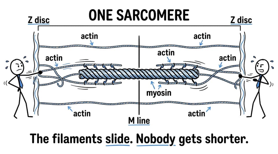
A sarcomere diagram represents the repeating contractile unit of a myofibril, extending from one Z disc to the next. For IB Biology HL, you should be able to identify its bands and filaments, explain how actin slides past myosin, and predict what changes during contraction. The key rule is simple: the sarcomere shortens, but the actin and myosin filaments do not.
Sarcomere diagram overview
Before exploring the mechanism, check that you can identify these features:
- Z discs: boundaries of one sarcomere
- Actin: thin filaments anchored to the Z discs
- Myosin: thick filaments occupying the centre
- M line: centre of the sarcomere
- I band: region containing actin but no myosin
- A band: entire length of the myosin filaments
- H zone: central region containing myosin but no actin overlap
- Titin: elastic protein associated with recoil and resistance to overstretching
This content appears in B3.3 Muscle and motility, an HL topic in the IB Biology course for first assessment in 2025. RevisionDojo's B3.3 Muscle and Motility resources bring the relevant Study Notes, Flashcards, lessons, and questions together.
How to label the structure correctly
Begin at the outside and work inward. The Z discs mark the boundaries, so a complete sarcomere always runs from Z disc to Z disc. Thin actin filaments extend inward from both boundaries. Thick myosin filaments occupy the centre and are organized around the M line.
The bands describe where those filaments are found:
- The I band contains thin filaments without overlapping myosin. A Z disc passes through its centre, so a diagram of one sarcomere usually shows half an I band at each end.
- The A band spans the entire length of the thick myosin filaments. Parts of it contain both actin and myosin.
- The H zone is the central part of the A band where there is myosin but no actin overlap in a relaxed sarcomere.
- The M line lies in the middle of the H zone and helps organize the thick filaments.
A common mistake is to label the A band as only the overlap region. It is not. Its width corresponds to the full length of myosin, including both the overlap regions and the H zone.
For a slower visual walkthrough, use RevisionDojo's sliding filament video lessons, then redraw the structure without looking.
What happens during muscle contraction
The sliding filament model explains why a sarcomere becomes shorter without either type of filament becoming shorter. Myosin heads form cross-bridges with exposed binding sites on actin. Their power strokes pull actin toward the M line, increasing the overlap between the thin and thick filaments.
As contraction proceeds:
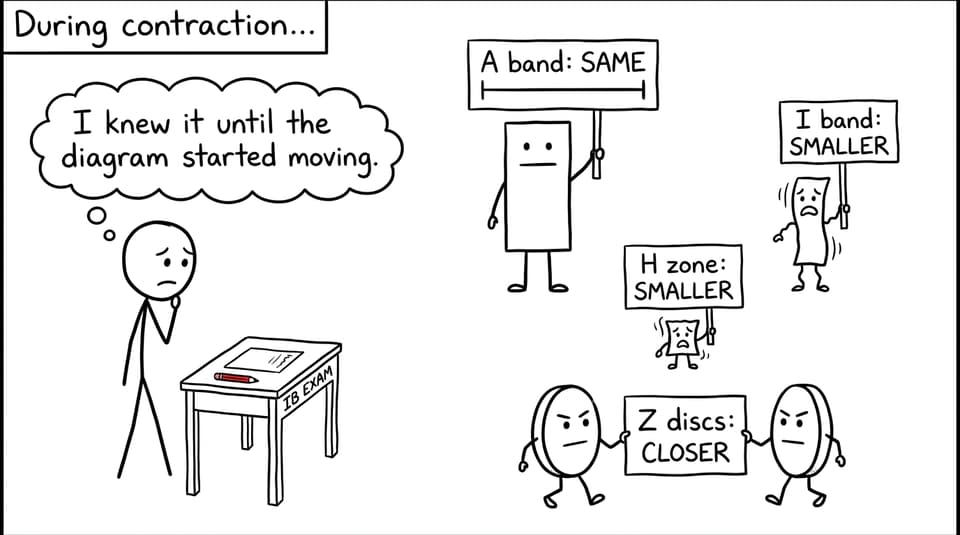
- Z discs move closer together.
- The sarcomere becomes shorter.
- The I bands become narrower.
- The H zone becomes narrower and may become difficult to see.
- Actin-myosin overlap increases.
- The A band remains the same width.
- Actin and myosin retain their original lengths.
Why does the A band remain constant? Because it represents the length of myosin, and myosin does not shorten. This is one of the most useful deductions you can make from the diagram.
The molecular sequence adds another layer. Calcium ions bind to troponin, causing a change that moves tropomyosin away from myosin-binding sites on actin. Myosin heads can then attach and perform power strokes. ATP binding allows myosin to detach from actin, while ATP hydrolysis helps reset the myosin head for another cycle.
Review this sequence in the sliding filament model notes or explore the connected explanation of actin and myosin in muscle contraction.
How sarcomere diagrams appear in IB exam questions
You may be asked to label a simplified diagram, interpret an electron micrograph, compare relaxed and contracted muscle, or explain observed changes. The challenge is often not recall alone. It is translating an unfamiliar image into a few stable structural rules.
Worked example
Question: A micrograph shows that the distance between adjacent Z discs decreases during contraction. Predict the changes in the I band, H zone and A band, and explain your answer.
Model answer: The I band becomes narrower because actin filaments slide farther over the myosin filaments. The H zone also becomes narrower because actin moves toward the centre of the sarcomere, reducing the region containing only myosin. The A band remains the same width because the myosin filaments do not change length. The Z discs move closer together, so the whole sarcomere shortens.
Notice the structure of that answer: observation, change, biological reason. It does not merely list what becomes smaller. It connects each visible change to filament movement.
Practise applying the same logic through the B3.3 Muscle and Motility Questionbank. RevisionDojo's Grading tools and AI Chat can then help identify vague language, missing links, or confusion between bands and filaments.
Common mistakes and misconceptions
Saying that actin or myosin contracts
The filaments slide; they do not shorten. It is the sarcomere, myofibril, muscle fibre, and eventually muscle that become shorter.
Drawing boundaries around the A band
One sarcomere extends from one Z disc to the next. The A band sits inside the sarcomere and is not its boundary.
Assuming every region changes width
The I band and H zone narrow, but the A band remains constant because myosin remains the same length.
Confusing the H zone with the overlap region
The H zone contains thick myosin filaments without actin overlap. It becomes smaller precisely because overlap increases during contraction.
Reversing troponin and tropomyosin
Calcium binds to troponin. The resulting conformational change shifts tropomyosin, exposing binding sites on actin.
Forgetting titin
The current syllabus also expects knowledge of titin. It contributes elastic recoil and helps prevent excessive stretching of the sarcomere. Consolidate that point with RevisionDojo's titin study notes.
Quick-reference summary
| Feature | Location or composition | Change during contraction |
|---|---|---|
| Z discs | Boundaries of a sarcomere | Move closer together |
| I band | Actin without myosin overlap | Becomes narrower |
| A band | Full length of myosin | Remains constant |
| H zone | Myosin without actin overlap | Becomes narrower |
| M line | Centre of the sarcomere | Remains central |
| Actin | Thin filament anchored to Z discs | Slides toward the M line |
| Myosin | Thick central filament | Retains its length |
| Sarcomere | Region from Z disc to Z disc | Shortens |
A strong final revision test is to draw a relaxed sarcomere, redraw it contracted, and explain every changed label aloud. Then use the IB Biology Questionbank to transfer that knowledge into data-based and short-answer questions.
Turn the diagram into marks
A sarcomere looks complicated because several labels occupy a small space. Yet the logic underneath is economical: boundaries move inward, overlap increases, and filament lengths remain constant. Once those relationships become secure, even an unfamiliar micrograph becomes manageable.
RevisionDojo is built for that transition from recognition to performance. Use its Study Notes and videos to understand the structure, Flashcards to retrieve each label, the Questionbank and Predicted Papers to practise application, and Mock Exams with Grading tools to sharpen your wording. If a mechanism still feels uncertain, AI Chat, the Coursework Library, and RevisionDojo Tutors provide additional routes to clarity.