Practice B3.3 Muscle and motility (HL) with authentic IB Biology exam questions for both SL and HL students. This question bank mirrors Paper 1A, 1B, 2 structure, covering key topics like cell biology, genetics, and ecology. Get instant solutions, detailed explanations, and build exam confidence with questions in the style of IB examiners.
| Sarcomere Region | Resting Length (μm) | Contracted Length (μm) |
|---|---|---|
| A-band | 1.8 | 1.8 |
| I-band | 0.7 | 0.4 |
| H-zone | 0.4 | 0.1 |
Based on the data, which conclusion best supports the sliding filament mechanism?
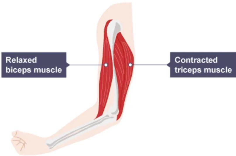
Refer carefully to the specific arrangement and position of the biceps and triceps in the image.

Which change would be observed in the diagram if the forearm were flexed instead of extended?
What is the role of calcium ions released from the sarcoplasmic reticulum?
What is the role of the joint capsule in an elbow joint?
What is a similarity between human and insect muscles?
Explain how the sliding filament model accounts for muscle contraction and relaxation including the roles of ATP and calcium ions.
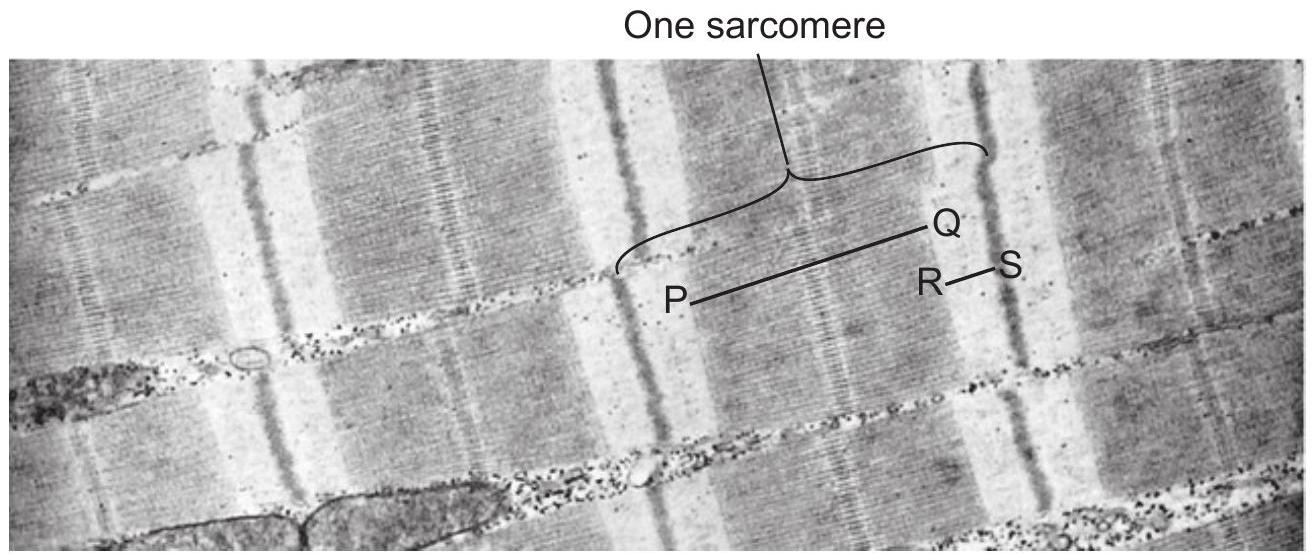
The electron micrograph shows sarcomeres in myofibrils of striated muscle during muscle contraction. The lines P-Q and R-S show two regions of one sarcomere.

How would regions P-Q and R-S change when the muscle relaxes?
| P-Q | R-S |
|---|---|
| wider | narrower |
| narrower | wider |
| wider | no change |
| no change | wider |
Which structure is used for locomotion in the earthworms but not in arthropods, bony fishes or birds?
A study measured the sarcomere length (µm) in isolated muscle fibers before and after ATP was added. The results are shown below:
| Condition | Sarcomere Length (µm) |
|---|---|
| Before ATP Addition | 2.5 |
| After ATP Addition | 2.1 |
Describe the change in sarcomere length after ATP addition and explain what this indicates about muscle contraction.
Explain the role of ATP in muscle contraction at the molecular level.
How does smooth muscle contraction differ from skeletal muscle contraction in terms of control?