

A phospholipid diagram should show a polar, hydrophilic phosphate-containing head connected through glycerol to two non-polar, hydrophobic fatty-acid tails. In a bilayer, the heads face the aqueous solutions on either side, while the tails point inward. That simple arrangement explains membrane formation, selective permeability, and several of the most testable ideas in IB DP Biology.
The drawing is small. The thinking behind it is not. A circle with two lines can represent amphipathic chemistry, spontaneous self-assembly, compartmentalization, and the boundary on which much of cell biology depends. The goal in an exam is therefore not artistic realism. It is biological precision.
Phospholipid diagram checklist
Before moving into the details, make sure you can do all of the following:
- Draw one phosphate-containing hydrophilic head.
- Add two fatty-acid tails, not one or three.
- Label the tails hydrophobic and non-polar.
- Use the term amphipathic correctly.
- Arrange phospholipids into two opposing layers when drawing a membrane.
- Show heads facing water and tails facing the bilayer's interior.
- Explain how the hydrophobic core restricts ions and most polar substances.
- Distinguish a phospholipid molecule from the complete fluid mosaic model.
These ideas sit across current syllabus content including B1.1 carbohydrates and lipids and B2.1 membranes and membrane transport. RevisionDojo's phospholipid bilayer formation notes and B2.1 membrane transport hub let you study both connections rather than memorizing the picture in isolation.
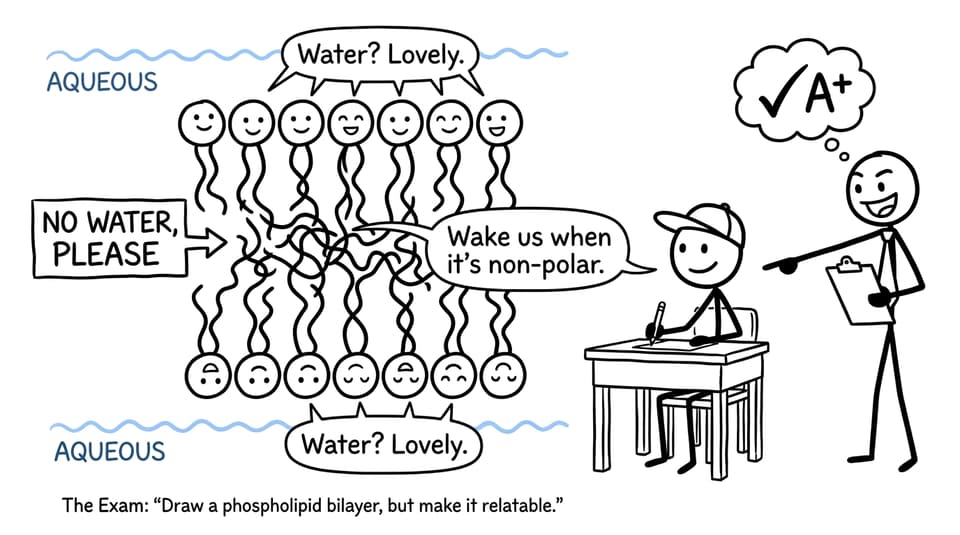
What each part of a phospholipid represents
A typical phospholipid contains glycerol, two fatty acids, and a phosphate-containing group. In simplified exam diagrams, the phosphate-containing region is represented by a circle and the fatty-acid chains by two lines. The glycerol connection may be shown separately in a molecular drawing, but it is often absorbed into the symbolic head when the question concerns membrane arrangement.
The hydrophilic head
The head is polar and interacts with water, so it is described as hydrophilic. In a plasma membrane, heads face the extracellular fluid on one surface and the aqueous cytosol on the other.
Writing only “phosphate” does not fully explain its behaviour. For an explanation question, connect the ideas:
The phosphate-containing head is polar, so it interacts with surrounding water and faces the aqueous environment.
The hydrophobic tails
The two fatty-acid chains are predominantly non-polar hydrocarbon regions. They are hydrophobic, meaning that they are excluded from favourable interactions with water. Inside a bilayer, the tails therefore cluster away from the aqueous solutions.
The tails are not attracted to water and do not face the cytosol. That orientation is one of the fastest ways to distinguish a correct diagram from an inverted one.
Why phospholipids are amphipathic
Amphipathic means possessing both hydrophilic and hydrophobic regions. This dual character causes phospholipids to form sheet-like bilayers spontaneously in aqueous conditions, with hydrophilic surfaces and a hydrophobic interior.
This is a cause-and-effect chain worth learning:
polar head plus non-polar tails -> amphipathic molecule -> bilayer formation in water -> hydrophobic membrane core -> selective barrier
For a focused visual lesson, use RevisionDojo's lipid bilayers study notes. The wider topic hub also includes explainer videos for students who learn more effectively by watching the arrangement develop step by step.
From one molecule to a phospholipid bilayer
A cell has aqueous fluid both inside and outside its plasma membrane. A stable membrane must therefore separate two watery environments without leaving hydrophobic tails exposed at either surface.
Phospholipids solve this through a bilayer:
- The outer row of heads faces extracellular fluid.
- The inner row of heads faces cytosol.
- The two sets of tails point toward one another.
- The membrane's centre becomes hydrophobic.
This is not merely a structural detail. The hydrophobic core has low permeability to ions and most polar molecules. Small non-polar molecules such as oxygen and carbon dioxide can move directly through the bilayer by simple diffusion, whereas ions generally require specific membrane proteins.
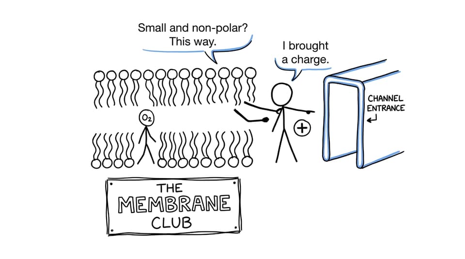
That distinction becomes clearer when you compare the diagram with RevisionDojo's guide to diffusion and active transport. The phospholipids and selective membranes guide develops the structure-function link further.
How phospholipid diagrams appear in IB Biology exams
The command word determines the required scope. “Draw a phospholipid” asks for one molecule. “Draw a phospholipid bilayer” requires two layers and correct orientation. “Draw the fluid mosaic model” demands more, including relevant membrane proteins and other requested components.
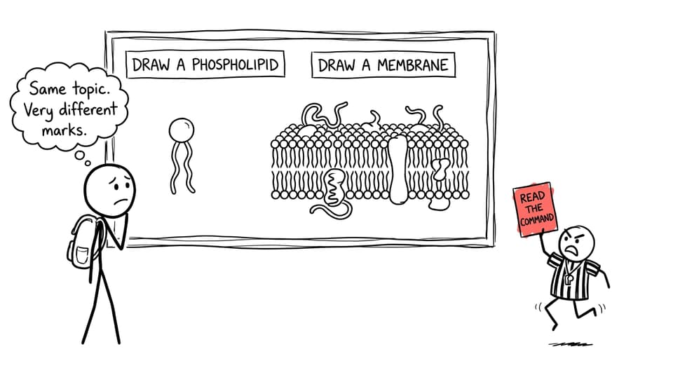
Worked example: draw and annotate a phospholipid
Possible prompt: Draw an annotated diagram of a phospholipid.
A strong response would include:
- One clearly defined head.
- Two separate tails attached to the head region.
- A label identifying the phosphate-containing head as polar and hydrophilic.
- A label identifying the fatty-acid tails as non-polar and hydrophobic.
- An annotation stating that the molecule is amphipathic.
Keep leader lines unambiguous. Do not let one label point vaguely toward the entire molecule when it is meant to identify a specific region.
Worked example: explain bilayer formation
Possible prompt: Explain why phospholipids form bilayers in water.
A concise answer could read:
Phospholipids are amphipathic. Their polar, hydrophilic heads interact with water, while their non-polar, hydrophobic tails orient away from it. Two layers form with the heads exposed to aqueous solutions and the tails facing inward. This shelters the tails from water and produces a stable hydrophobic core.
Notice the progression: property, orientation, arrangement, consequence. Naming the head and tails without explaining their behaviour would not fully answer “explain.”
Worked example: connect structure to permeability
Possible prompt: Explain why an ion cannot easily diffuse through the phospholipid bilayer.
A strong response is:
An ion is charged and hydrophilic, whereas the centre of the bilayer is formed by non-polar, hydrophobic fatty-acid tails. The ion cannot pass readily through this hydrophobic core, so movement normally depends on a suitable channel or transport protein.
Once your explanation is secure, apply it through the B2.1 membrane Questionbank. Retrieval practice through the matching membrane transport flashcards can help make terms such as amphipathic, polar, and hydrophobic automatic.
Drawing the fluid mosaic model
A phospholipid diagram is not automatically a complete membrane model. If the question asks for a two-dimensional representation of the fluid mosaic model, the current IB Biology guide expects students to understand a broader arrangement that can include:
- A phospholipid bilayer with hydrophilic and hydrophobic regions indicated.
- Integral proteins embedded in or spanning the bilayer.
- Peripheral proteins associated with a membrane surface.
- Glycoproteins with carbohydrate chains projecting from the extracellular side.
- Cholesterol positioned among phospholipid tails in animal membranes.
The term fluid refers to the capacity of membrane components, including phospholipids, to move laterally. Mosaic describes the varied proteins and other components distributed through or associated with the bilayer. Review the complete structure with RevisionDojo's fluid mosaic model resources.
Common mistakes that lose marks
Reversing the orientation
Heads must face the aqueous environments; tails must face inward. Writing correct labels on an inverted bilayer does not repair the biological error.
Drawing a single membrane layer
A plasma membrane is based on a bilayer, meaning two phospholipid layers. A monolayer does not separate two aqueous compartments in the required arrangement.
Giving each phospholipid one tail
The standard IB representation has two fatty-acid tails. Make both visible, even when drawing quickly.
Confusing hydrophobic with impermeable
The hydrophobic core restricts ions and most polar molecules, but the membrane is not completely impermeable. Small non-polar molecules can cross directly, and proteins provide selective routes for other substances.
Adding every membrane component to every question
Extra detail can consume time and make a drawing harder to interpret. Match the requested object: molecule, bilayer, or fluid mosaic model.
Using labels without explanations
“Head” and “tail” identify structures. “Polar and hydrophilic” and “non-polar and hydrophobic” explain chemical properties. For explanation questions, connect those properties to orientation, bilayer formation, or permeability.
Quick-reference summary
| Feature | What to show or state |
|---|---|
| Head | Phosphate-containing, polar, hydrophilic |
| Tails | Two fatty-acid chains, non-polar, hydrophobic |
| Whole molecule | Amphipathic |
| Bilayer surfaces | Heads face extracellular fluid and cytosol |
| Bilayer centre | Tails face inward, forming a hydrophobic core |
| Membrane consequence | Low permeability to ions and most polar molecules |
| Simple diffusion | Small non-polar molecules can cross directly |
| Full membrane model | Add integral and peripheral proteins, glycoproteins, and cholesterol where relevant |
Turn the drawing into exam-ready understanding
The most useful way to revise this topic is to move through three levels. First, draw and label one phospholipid from memory. Next, build it into a bilayer and explain why the arrangement forms. Finally, use that structure to predict which substances can cross and which require proteins.
RevisionDojo brings that sequence together. Use syllabus-aligned Study Notes and Flashcards to establish the language, then answer targeted questions in the Questionbank and use AI Chat or Grading tools to diagnose imprecise explanations. As exams approach, Predicted Papers and Mock Exams can place membrane questions under realistic time pressure, while the Coursework Library and Tutors support the wider demands of IB Biology.
A good drawing does more than resemble a membrane. It shows that you understand why the membrane exists in that form. Once every line represents a reason, the diagram becomes much easier to remember and much harder to mislabel.