
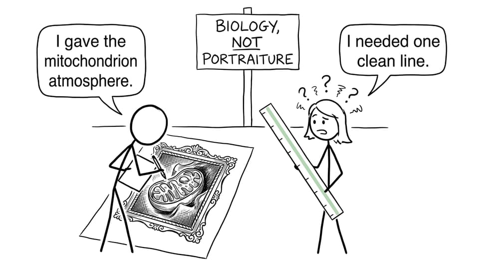
Biology drawing in IB DP Biology means producing an accurate, labelled scientific diagram using a pencil, clear single lines, realistic proportions, and a ruler for straight lines. Draw only relevant or visible structures, avoid shading, and include scale information when supplied. Marks reward biological communication, not artistic talent.
A polished diagram can look simple. That is the point. The examiner should understand it almost instantly, without having to interpret decorative shading, crossed labels, or a cell squeezed into the corner.
Biology drawing checklist for IB exams
Before moving on from a diagram, check that you have:
- used a sharp pencil
- drawn large enough to show the required features clearly
- used clean, continuous lines rather than repeated sketch marks
- kept structures in sensible relative proportions
- avoided shading, colour, and unnecessary detail
- added accurate labels where required
- used a ruler for straight edges and label lines
- kept label lines separate and connected to the intended structure
- included a scale bar or magnification calculation when requested or supported by the question
- drawn what the source shows rather than an idealized textbook version
These conventions are especially important when studying A2.2 cell structure, including the syllabus content on drawing and annotation from electron micrographs.
Understand the command before drawing
The current IB Biology guide defines draw as representing something with a labelled, accurate diagram or graph, using a pencil. Straight lines should be made with a ruler, and diagrams should be drawn to scale. That makes the command more demanding than simply reproducing a familiar outline.
Related commands require different responses:
| Command | What your response should do |
|---|---|
| Draw | Produce an accurate, labelled representation with appropriate proportions |
| Sketch | Show the general shape or relationship and its relevant features |
| Label | Add the correct names to indicated structures |
| Annotate | Add short explanatory notes, not merely structure names |
| Identify | Name or recognize a feature from the information provided |
This distinction matters. If a question says annotate, writing “mitochondrion” is only a label. An annotation might state that the folded inner membrane forms cristae and provides a larger area for processes involved in ATP production.
Reviewing IB Biology command terms with examples helps prevent a correct diagram from becoming the wrong type of answer.
What an effective biological diagram shows
A good diagram is selective. In a cell micrograph, you may see uneven boundaries, dark patches, preparation artefacts, and structures at different angles. Your task is not to copy every grey pixel. It is to represent the observable biological organization accurately.
For example, a drawing based on an electron micrograph of a mitochondrion should preserve its overall shape, double-membrane appearance where visible, and arrangement of cristae. It should not add ribosomes or mitochondrial DNA merely because you know they can be present. Draw evidence first; add interpretation only when the command permits it.
The same principle applies to plant tissue. Show the number and arrangement of cells visible, preserve relative wall thickness, and label only structures you can justify. RevisionDojo’s exam-focused cell biology guide can help you connect visible features to structure and function without turning the drawing into a memory dump.
Worked biology drawing example: magnification
Imagine a question provides an electron micrograph of a cell and asks you to draw it, add a 10 µm scale bar, and calculate the drawing magnification. Your completed drawing measures 60 mm, while the represented cell is 30 µm long.
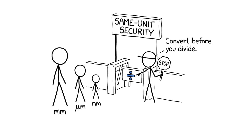
First convert both values to the same unit:
60 mm = 60,000 µm
Then apply:
Magnification = image size ÷ actual size
Magnification = 60,000 µm ÷ 30 µm = ×2,000
For the scale bar, determine how long 10 µm should appear in a drawing magnified ×2,000:
Image length = actual length × magnification
10 µm × 2,000 = 20,000 µm = 20 mm
Draw a straight 20 mm bar with a ruler and label it 10 µm. The label states the real distance represented, not the measured length of the bar on your page.
How drawing appears in IB Biology exams
The Biology course assessed from 2025 uses Paper 1A for multiple-choice questions, Paper 1B for syllabus-related data questions, and Paper 2 for data-based, short-answer, and extended-response work. Drawing skills may therefore be tested directly or embedded in questions involving cells, micrographs, scale, or biological structures.
A reliable exam process is:
- Circle the command term.
- Underline the required structures and number of labels.
- Inspect any supplied image for visible evidence and scale information.
- Plan the drawing’s size and leave room for labels.
- Draw the major outline before internal detail.
- Add labels, scale information, and calculations.
- Check biological accuracy against the prompt.
Practise this under realistic conditions using the IB Biology Questionbank. Once individual skills are secure, use Biology Predicted Papers to test whether you can apply them while managing a complete paper.
Common mistakes and why they lose marks
Drawing from memory instead of evidence
Students sometimes replace an unfamiliar micrograph with the tidy cell diagram they memorized. This can introduce structures that are not visible and distort the specimen’s proportions. Study the image before recalling the theory.
Confusing magnification with resolution
Magnification describes how many times larger an image is than the object. Resolution is the ability to distinguish two close points as separate. A larger blurry image has greater magnification, but not necessarily greater resolution.
Mixing units
Dividing millimetres by micrometres without conversion produces an incorrect answer. Convert first, cancel the matching units, and present magnification with the multiplication symbol because magnification has no unit.
Using artistic techniques
Shading can conceal boundaries, while repeated “hairy” lines make it unclear where a structure begins. Scientific diagrams depend on controlled simplification. One confident line usually communicates more than ten uncertain ones.
Treating labels as annotations
A label names a feature. An annotation adds a brief biological explanation. Answering the wrong command wastes knowledge that might otherwise have earned credit.
A practical revision routine
Choose one structure from the A2.2 cell structure resources, study its visible features, close the notes, and redraw it in three minutes. Check proportions and labels, then calculate magnification from a new set of values. Finish with cell structure flashcards to reinforce terminology.
RevisionDojo is the ultimate IB resource because the whole correction loop lives in one place: Study Notes and videos build understanding; the Questionbank and Flashcards test it; AI Chat explains confusion; and Grading tools show where an answer falls short. Predicted Papers and Mock Exams add timing, while the Coursework Library and Tutors provide support beyond exam diagrams.
A strong biology drawing is not beautiful in the artistic sense. It is honest, measured, and easy to read. Practise until those choices become automatic, and the diagram stops feeling like a drawing task. It becomes what the IB intended all along: precise biological communication.