


An action potential graph plots membrane potential against time as a neuron depolarizes, repolarizes, briefly hyperpolarizes, and returns to resting potential. The rising phase results mainly from Na⁺ entering through voltage-gated sodium channels; the falling phase results mainly from K⁺ leaving through voltage-gated potassium channels.
At first glance, the trace looks like a narrow mountain followed by a small dip. But every slope represents a change in membrane permeability, ion movement, and channel state. Once you can translate shape into mechanism, the graph becomes one of the more predictable parts of IB Biology neural signalling.
What IB Biology students need to know
The current IB Biology course places neural signalling in C2.2. All students need the principles of resting potentials and nerve impulses, while HL students study depolarization, repolarization, action-potential propagation, and oscilloscope traces in greater depth.
Use this checklist when revising:
- Identify the x-axis as time and the y-axis as membrane potential in millivolts.
- Recognize resting potential, threshold, depolarization, repolarization, and hyperpolarization.
- Link the rising phase to Na⁺ influx.
- Link the falling phase to K⁺ efflux.
- Explain the all-or-nothing principle and refractory period.
- Read exact values from the trace rather than assuming every neuron has identical voltages.
For the wider syllabus context, start with C2.2 Neural Signalling study notes or review the complete C2.2 Neural Signalling topic hub.
Reading an action potential graph phase by phase
Typical teaching diagrams use approximately -70 mV for resting potential, around -55 mV for threshold, and roughly +30 to +40 mV for the peak. These are useful reference values, not universal constants. In an exam, prioritize the numbers shown on the supplied graph.
| Graph region | What is happening |
|---|---|
| Resting potential | The inside of the neuron is negative relative to the outside; ion gradients and selective membrane permeability maintain polarization. |
| Threshold | Sufficient depolarization triggers the opening of voltage-gated Na⁺ channels. |
| Depolarization | Na⁺ enters down its electrochemical gradient, making the inside less negative and then positive. |
| Peak | Na⁺ channels inactivate while delayed voltage-gated K⁺ channels are open. |
| Repolarization | K⁺ leaves the neuron, causing the membrane potential to become negative again. |
| Hyperpolarization | K⁺ channels close slowly, so continued K⁺ efflux takes the potential below its resting level. |
| Return to rest | Channel states reset, while the sodium-potassium pump and leak channels maintain the longer-term ion gradients. |
Resting potential is not inactivity
A resting neuron is electrically polarized, not switched off. The sodium-potassium pump uses ATP to transport Na⁺ out and K⁺ in, helping establish their concentration gradients. The membrane is also more permeable to K⁺ than Na⁺ at rest, contributing to the negative internal potential.
A common weak answer says only that the pump “makes the inside negative.” A stronger answer connects active transport, unequal ion distributions, selective permeability, and the resulting potential difference.
Threshold and depolarization
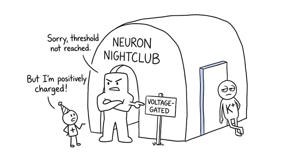
A stimulus initially makes the membrane potential less negative. If threshold is reached, voltage-gated sodium channels open. Na⁺ enters by facilitated diffusion down its electrochemical gradient, which causes further depolarization and opens more sodium channels.
This positive feedback produces the steep upward line. If threshold is not reached, a full action potential is not generated. Once it is reached, increasing stimulus strength does not create a taller action potential; stronger stimulation is generally represented by a higher frequency of impulses.
Repolarization and hyperpolarization
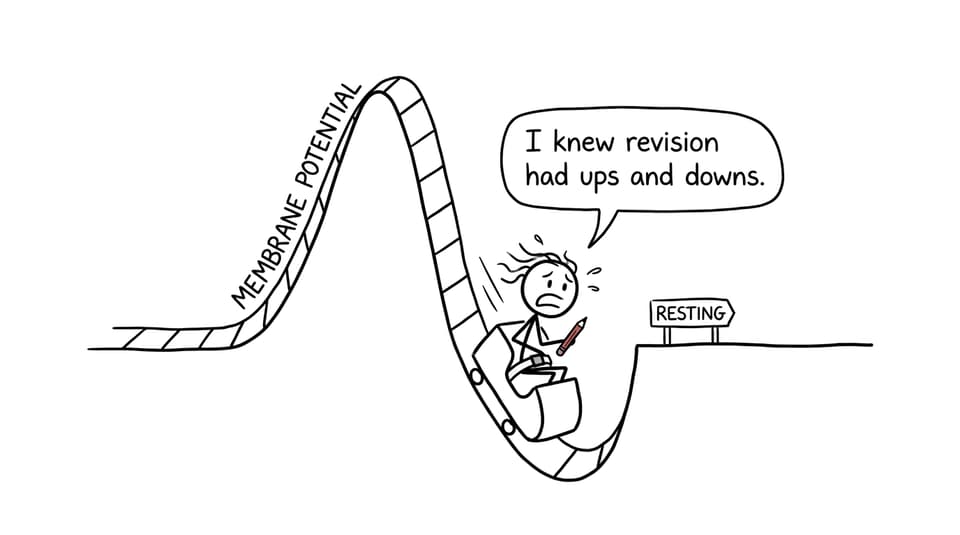
Near the peak, voltage-gated sodium channels inactivate and voltage-gated potassium channels remain open. K⁺ moves out, taking positive charge with it. The membrane potential therefore falls toward its resting value.
Potassium channels close with a delay, so K⁺ may continue leaving after the resting potential has been crossed. This produces the undershoot called hyperpolarization. The channel sequence matters, so reinforce it with RevisionDojo’s gated ion channel notes.
The refractory period
During the absolute refractory period, inactivated sodium channels cannot immediately reopen. During the relative refractory period, another action potential is possible, but stronger stimulation may be required because the membrane is hyperpolarized and some channels have not fully reset.
Refractoriness limits firing frequency and helps prevent immediate backward propagation. To connect one trace with movement along an axon, review how local currents propagate an action potential.
Worked action potential graph example
Consider a hypothetical trace with these readings:
- Resting potential: -70 mV
- Threshold: -55 mV
- Peak: +30 mV
- Lowest point during hyperpolarization: -80 mV
Question: Calculate the change in membrane potential from rest to the peak and explain the rising phase.
Calculation:
+30 mV - (-70 mV) = 100 mV increase
Model explanation:
The membrane reaches threshold, causing voltage-gated sodium channels to open. Na⁺ enters the neuron down its electrochemical gradient. The influx of positive ions makes the inside increasingly positive, producing rapid depolarization from -70 mV to +30 mV.
Notice the division of labour. The calculation reports magnitude with units; the explanation names the channel, ion, direction, transport mechanism, and electrical consequence.
Exam questions may also ask you to interpret an oscilloscope trace, compare two neurons, or predict the effect of blocking an ion channel. Use the C2.2 Neural Signalling Questionbank to practise these variations, then watch the C2.2 neural signalling video lessons when a moving explanation would help.
How to answer graph questions in an exam
Follow a three-step sequence:
- Describe the evidence. Quote the direction, values, and units shown.
- Identify the phase. Name depolarization, repolarization, or hyperpolarization.
- Explain the mechanism. State which channels open or inactivate, which ion moves, and in which direction.
For example, do not write only, “The voltage goes down.” Write: “The membrane potential decreases from +30 mV toward -70 mV as voltage-gated K⁺ channels open and K⁺ diffuses out of the neuron.”
Command terms also matter. State requires a short answer; describe requires the observed pattern; explain requires a biological cause. The broader IB Biology animal physiology guide shows how to build these cause-and-effect responses.
Common mistakes that lose marks
- Reversing ion directions: Na⁺ enters during depolarization; K⁺ leaves during repolarization.
- Saying ions move by active transport during the spike: movement through open channels is passive, down electrochemical gradients.
- Giving the pump all the credit for repolarization: rapid repolarization is primarily caused by K⁺ efflux through voltage-gated channels.
- Calling hyperpolarization depolarization: hyperpolarization makes the inside more negative than at rest.
- Claiming stronger stimuli make taller spikes: action potentials are all-or-nothing once threshold is reached.
- Ignoring graph data: use the values provided rather than forcing memorized textbook values onto the trace.
- Omitting units: membrane potential is measured in mV and time may be measured in ms.
A quick revision routine
Draw the trace from memory, label every phase, and add one ion movement beneath each slope. Then cover it and redraw it in under two minutes. Finish with a small set of questions from the IB Biology Questionbank, checking whether each error came from knowledge, graph reading, or wording.
RevisionDojo turns that routine into a complete loop: learn with Study Notes, retrieve with Flashcards, clarify with AI Chat, and apply with the Questionbank. Grading tools, Predicted Papers, and Mock Exams help build exam precision, while the Coursework Library and Tutors support the wider IB journey. The graph may last only milliseconds, but understanding it can connect membranes, transport, neurons, and exam technique in one clear picture.