


If you have ever copied a long paragraph by hand, you know the weird moment where your brain starts to autocomplete words that are not actually there. DNA replication has the same problem: speed invites mistakes. Yet cells copy billions of bases with astonishing accuracy.
A quiet reason sits inside the double helix itself: antiparallel DNA. For IB Biology students, this is one of those details that looks like a diagram-labeling exercise until you realize it is the rule that makes the whole system workable.
The IB Biology checklist: what to remember in 60 seconds
Use this quick list before you open a markscheme:
-
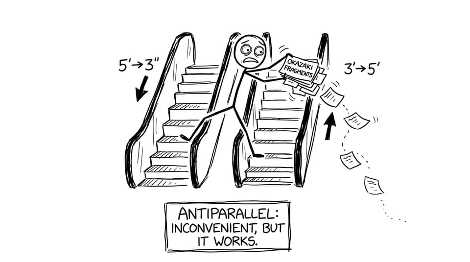
Antiparallel means one strand runs 5' to 3' and the other runs 3' to 5'.
-
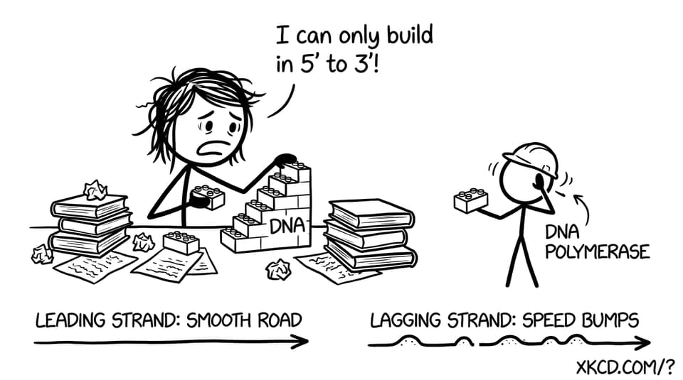
DNA polymerase adds nucleotides only to a free 3' OH, so new DNA is made 5' to 3'.
-
This creates a leading strand (continuous) and a lagging strand (discontinuous, via Okazaki fragments).
-
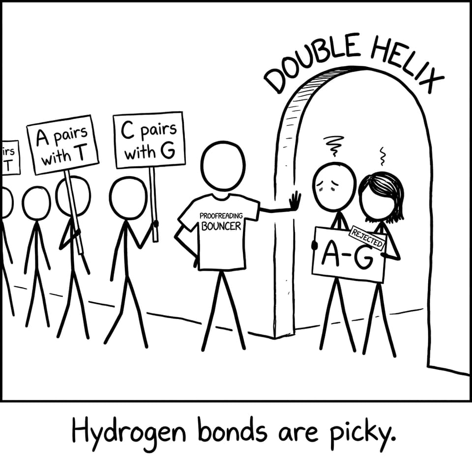
Correct complementary base pairing (A-T, C-G) fits the helix geometry, helping accuracy.
-
Proofreading and repair work better when the helix has a consistent, predictable shape.
If you want the syllabus-aligned version of the whole pathway, start with D1.1 DNA Replication and then drill specifics in IB Biology style.
Why antiparallel DNA matters for replication accuracy
In IB Biology, the phrase “DNA polymerase works 5' to 3'” shows up everywhere. The deeper point is chemical: polymerase needs that 3' hydroxyl group to attach the next nucleotide. There is no workaround in normal replication.
Now connect that to antiparallel strands. Because the templates point in opposite directions, the replication fork must solve a coordination problem: copying both templates while staying loyal to the 5' to 3' rule.
That coordination is not just about speed. It is about accuracy. When the process is organized and predictable, the enzyme can detect “wrong fits” faster and fix them with fewer side effects.
For a clean, exam-ready walkthrough, revise the topic page D1.1.1 DNA replication alongside this concept.
Leading vs lagging strand: the elegant compromise
The antiparallel layout forces the fork to build:
-
Leading strand: synthesized continuously in the same overall direction as fork movement.
-
Lagging strand: synthesized discontinuously, away from the fork, in short Okazaki fragments that later get joined.
This is the part many IB Biology students memorize, but you score higher when you explain why it must be this way: polymerase cannot flip direction, and templates are opposite.
RevisionDojo’s notes make this comparison especially crisp: Differences between replication on the leading and lagging strand.
Antiparallel structure improves “error visibility”
Replication accuracy is not only about picking the right base. It is about noticing when something looks wrong.
Correct base pairs create consistent spacing and hydrogen-bond geometry. A mismatch subtly warps the helix, like a zipper tooth bent out of shape. That distortion is a physical signal that something has gone off-script.
In IB Biology, you can describe this as “incorrect pairing disrupts helix geometry,” which helps polymerase proofreading and downstream repair systems identify errors.
To strengthen your explanation of why the template system is so reliable, connect antiparallel DNA to the bigger principle of fidelity in Semi-conservative nature of DNA replication.
Proofreading works because directionality is consistent
Polymerase proofreading (often discussed as a built-in check) depends on stable, repeatable alignment between:
-
the template base,
-
the incoming nucleotide,
-
and the active site of the enzyme.
Antiparallel DNA gives enzymes a consistent road map: templates are read in a predictable direction, and synthesis always proceeds 5' to 3'. If strands were parallel, base pairing would not align correctly and the entire “fit-check-correct” rhythm would become unreliable.
If you want to revise the full cast of enzymes that keep this process accurate, use 2.7 DNA replication, transcription and translation Notes and then zoom into enzyme roles with D1.1.8 DNA primase, DNA polymerase I, DNA polymerase III and DNA ligase.
How to turn this into exam marks (IB Biology phrasing)
A high-scoring IB Biology explanation usually includes:
-
Define antiparallel using 5' and 3' ends.
-
State polymerase limitation: adds only to 3' OH, so synthesis is 5' to 3'.
-
Explain consequence: leading continuous, lagging discontinuous with Okazaki fragments.
-
Link to accuracy: correct base pairing stabilizes helix; mismatches distort structure; proofreading is more effective.
Then practice it under time pressure using RevisionDojo’s Questionbank. A good starting point for this sub-topic is A1.2 Nucleic acids Questionbank.
Bring it home with RevisionDojo
Antiparallel DNA is a small structural detail with big consequences: it makes directionality predictable, coordination possible, and mistakes easier to spot. That is exactly the kind of structure-to-function link that IB Biology examiners reward.
When you are ready to turn understanding into marks, use RevisionDojo as your full stack: Study Notes for clarity, Flashcards for active recall, the Questionbank for exam-style practice, AI Chat when you get stuck mid-explanation, Grading tools to tighten wording, plus Predicted Papers, Mock Exams, the Coursework Library, and Tutors when you want your revision to feel guided rather than improvised. Start your next session from the IB Biology resources hub and keep IB Biology replication accuracy on your side.